| Diagnostic imaging, CT scan, Egyptology | |
Results of Tutankhamen scan revealed9 March 2005 The mummy of Tutankhamen was discovered in Egypt's Valley of Kings in 1922. An initial X-ray analysis in 1968 revealed a bone splinter embedded in the pharaoh's skull. This fact — coupled with the body's obviously hasty mummification and burial — led to speculation that Tutankhamen had died from head injuries, and possibly been murdered. The now completed analysis of the CT examination, based on images generated from a total of 1,700 slices, found no evidence for this theory. But the Pharaoh may have suffered from a broken thigh shortly before his death at the age of 19. Some members of the examination team say that the Pharaoh may have died from an infection of this wound. They refer to the fact that the CT images revealed embalming resin inside the wound, and that there was no sign of a healing process. Other scientists on the team doubt that the injury was the cause of the king's death. They believe the wound could have been inflicted later by archaeologists examining the mummy, arguing that there was no evidence of haematoma, which should be there if the injury was inflicted during the Pharaoh's lifetime. This examination is part of a research project being conducted by Egypt's Supreme Council of Antiquities. The project also includes meticulous CT scans of a large number of other Egyptian mummies. To support the project, Siemens has provided a special CT system, which is installed on a trailer — making it transportable to wherever it is needed. With this device, the fragile remains of Egypt's ancient people can be studied with a minimum of movement and disturbance.
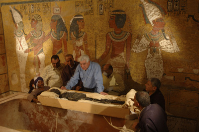
Inside King Tutankhamun’s tomb, Zahi Hawass, head of the Egyptian Supreme
Council of Antiquities, and a team of Egyptian researchers examine the
3,300-year-old mummy as it is removed from its sarcophagus prior to being
CT-scanned on Jan. 5, 2005.
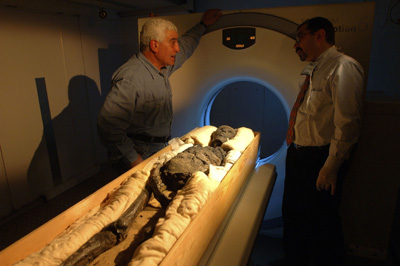
Zahi Hawass (left), head of the Egyptian Supreme Council of Antiquities,
looks on as the 3,300-year-old mummy of King Tutankhamun is prepared for
scanning. The CT scan took place on Jan. 5, 2005, outside Tutankhamun's tomb
in the Valley of the Kings and will reveal new information about this
enigmatic king. See also CT scans unwrap secrets of British Museum's Egyptian mummies CT scans and DNA tests help unveil mystery of long-lost female pharaoh |