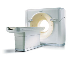
Philips 64-slice CT scanner installed in University of Chicago Hospitals
1 April 2005
The second 64-slice computed tomography scanner ever produced by Philips
Medical Imaging, and the first to reach the United States, has been
installed and is now in clinical use at the University of Chicago Hospitals.
The scanner, which has four times as many detectors as a typical
multi-detector CT scanner, combines unrivaled image quality with remarkable
speed. It can produce detailed pictures of any organ in a few seconds and
provide sharp, clear, three-dimensional images, including 3-D views of the
blood vessels, in an instant.
 "The
technology is stunning," said Michael Vannier, M.D., professor of radiology
at the University of Chicago. "We can perform much more detailed analysis of
very complex anatomy with a speed and confidence that we couldn't aspire to
a year ago. It has changed what we can look for and how we look at it when
we find it." "The
technology is stunning," said Michael Vannier, M.D., professor of radiology
at the University of Chicago. "We can perform much more detailed analysis of
very complex anatomy with a speed and confidence that we couldn't aspire to
a year ago. It has changed what we can look for and how we look at it when
we find it."
A complete chest scan used to include about 100 to 200 slices and take about
30 minutes, Vannier said. "Now we typically collect ten times as many slices
in less than half the time."
It has also changed the way radiologists look at CT scans. Prior to the
introduction of multi-detector CT scanners, doctors examined individual
slices. "Now we look at reconstructed three-dimensional views," Vannier
said, "and the doctors who send us patients insist on reconstructions. They
won't tolerate a collection of slices anymore."
"This new generation of scanners is going to revolutionize the way we detect
and diagnose heart disease," said Dianna Bardo, M.D., assistant professor of
radiology at the University of Chicago. "We have already become reliant upon
to these images in working up children with congenital heart disease, and
multi-detector CT scans of the heart are increasingly being used instead of
angiograms to detect clogged arteries in older patients. We're still
learning what this technology can do."
Last spring, the University of Chicago Hospitals entered a partnership with
Philips to serve as a clinical testing and development site for Philips'
newest equipment. The arrangement brings Philips' most advanced imaging
technologies to the University of Chicago. In exchange, the University's
renowned experts in computer detection and diagnosis help Philips test and
improve their image-processing software.
Last summer, Philips installed four new 16-slice scanners at the Hospitals
as well as one of their first 40-slice CT scanners, at the time one of eight
such devices in the world. In March 2005 they brought in the 64-slice
scanner.
Other companies, including General Electric, Siemens and Toshiba are
competing to produce and distribute similar multi-detector CT scanners,
which combine the highest possible image resolution with breathtaking speed.
Many standard scanners have only two, four, six or ten detectors. One year
ago a top-of-the-line scanner might have 16.
Each detector picks up an x-ray beam as it spins around the body and then
computes the densities of the tissues that beam has passed through to
produce a thin image of that narrow slice of the body.
The more detectors a scanner has, the closer they can be packed together,
which improves resolution, and the faster they can gather slices, which
increases speed. The computer then collects the images, stacks up the slices
like a loaf of bread with each slice thinner than a penny, and presents a
three-dimensional picture of the inside of the body. Software allows the
radiologist to adjust the images to highlight specific tissues.
The multi-detector scanners can be packed less than a millimeter apart and
take less than half a second to circle the body. So a 16-slice scanner can
cover 8 to 12 millimeters in one pass or about an inch a second. A 40-slice
scanner collects images covering 20 to 32 millimeters in a single pass and a
tightly packed 64-slice device can cover about 40 millimeters at a pass,
which takes 0.4 seconds.
At that rate, a 64-slice scanner can gather a high-resolution image of a
heart, brain or a pair of lungs in about five seconds. A scan of the whole
body, ( in search of a blood clot, for example, that has become a source of
emboli ) takes about 30 seconds.
The technology has been particularly exciting for studying the beating
heart, providing the first clear non-invasive images of the heart and its
major vessels. The scans can be timed to use only images gathered between
contractions, so that the heart and its vessels can be seen without the
blurring caused by motion.
In fact, these scanners have already provoked "turf" battles between
radiologists and the cardiologists who perform diagnostic angiograms --
until now the gold standard for assessing the coronary arteries. The best
multi-detector CT scans can rival angiography for detail and are quicker,
more convenient, less expensive and safer than an angiogram, as well as
exposing the patient to less radiation.
"We've already switched from catheter-based to CT-based imaging in the
brain," said Bardo. "The heart may come next."
"We are still sorting this issue out," cautions David Faxon, M.D., chief of
cardiology at the University of Chicago and past president of the American
Heart Association. "CT images have improved dramatically in the last few
years," he said, "but there are still certain patients for whom angiography
is more informative, especially for the small and distal vessels. I am sure,
however, that the detail and resolution of these images will improve with
time."
"What is clear right now," Faxon added, "is that when they combine tools and
experience, radiologists and cardiologists can now look at the precise
anatomy of the heart and its vessels in ways we only dreamed of not all that
long ago."
The scanners are beginning to have an impact on cancer diagnosis and
treatment as well. Nearly 60 percent of CT scans at the University of
Chicago Hospitals are done for cancer. The speed and precision of these new
scanners not only improves the image quality, but also "lets us look at
dynamic processes," Vannier explained. "Instead of just monitoring changes
in tumor size, we can watch the perfusion of a contrast agent as it moves
toward, around and through a tumor," he said. "This can provide an early
view of how a patient is responding to therapy. It helps us predict, rather
than simply describe responses to treatment."
Other promising indications for multi-slice scanners include evaluation of
plaque within the carotid arteries ( 5 to 8 seconds ), searching for
pulmonary emboli ( 5 seconds, less than an easy breath hold ), coronary
artery imaging ( 10 seconds, including distal segments and multiple arterial
branches ).
The scans have their own limitations. Although the scanner table is built to
support up to 450 pounds, it can be difficult to accommodate patients who
are morbidly obese. Each scanner costs between $1.5 million to $2 million.
One remaining challenge is image storage. The Philips "Brilliance
Workstation" collects and processes the images within seconds but the
enormous files can overwhelm the computers that store them.
"We never used to gather so much information so quickly," points out
Vannier. "We currently perform about 4,000 CT scans a month, each including
1,000 to 2,000 discreet slices, and those numbers are increasing. We have to
rethink how we can compress and store so much data, yet retain easy access
to these images when we need them."
To top |
|