| Laboratory systems, information technology | |
Leica microsystems launches networked imaging solution for histopathologists9 October 2006 Wetzlar, Germany. Leica Microsystems has designed a new networked imaging solution for the histopathology laboratory that makes it easier to view, store and share images of specimens.
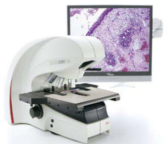
The Leica DMD108 system provides high-quality images directly on a monitor using a high-resolution camera and powerful image processing software. It generates high-resolution images with brilliant colour fidelity that equal those produced using a conventional microscope. Histopathologists can use the Leica DMD108 to
photograph specimen details of interest or compare tissue sections. Images
are easily stored with the click of a mouse and can be retrieved at any
time. Size ratios are also calculated by a simple keystroke. The
histopathologist can then audio-record the diagnosis directly onto The system
provides an ideal link between pathologists around the world. Now, holding
live discussions about specific specimens is amazingly easy with Leica’s new
network imaging solution, and it is
|
 The
Leica DMD108 offers an innovative solution for histopathologists that
increases physical comfort, significantly speeds daily workflow without
changing the process, and provides an easy-to-use network solution for
sharing data. The solution has been extensively tested with pathologists to
ensure that their daily requirements are met.
The
Leica DMD108 offers an innovative solution for histopathologists that
increases physical comfort, significantly speeds daily workflow without
changing the process, and provides an easy-to-use network solution for
sharing data. The solution has been extensively tested with pathologists to
ensure that their daily requirements are met.