Laser fluorescence technology highlights cell surface protein receptors in real time
9 September 2008
Researchers at The University of Nottingham have developed laser fluorescence technology that enables observation of protein activity on living cell surfaces for the first time.
The cutting-edge technology has helped to attract a £1.3 million grant from the UK Medical Research Council for a five-year project that will offer new insights into chemical activity taking place within single cells. It could contribute to the design of new drugs to treat human inflammatory diseases such as asthma and arthritis with fewer side effects.
The team, involving Professor Steve Hill and Dr Steve Briddon from the University’s School of Biomedical Science and Dr Barrie Kellam from the School of Pharmacy, is concentrating on a type of specialised docking site (receptor) on the surface of a cell that recognises and responds to a natural chemical within the body called adenosine.
These A3-adenosine receptors work within the body by binding with proteins to cause a response within cells and are found in very tiny and highly specialised area of a cell membrane called microdomains. Microdomains contain a collection of different molecules that are involved in telling the cell how to respond to drugs or hormones.
It is believed that these receptors play an important role in inflammation within the body and knowing more about how they operate could inform the future development of anti-inflammatory drugs that target just those receptors in the relevant microdomain of the cell, without influencing the same receptors in other areas of the cell. However, scientists have never before been able to look in detail at their activity within these tiny microscopic regions of a living cell.
The Nottingham researchers have solved this problem by creating novel drug molecules which have fluorescent labels attached. Using a cutting edge laser technology called fluorescence correlation spectroscopy, the fluorescent drug molecules can be detected as they glow under the laser beam of a highly sensitive microscope. This allows their binding to the receptor to be followed for the first time in real time at the single molecule level.
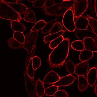
A fluorescent drug (in red) binding to an
adenosine receptor on the surface of living cells.
Leading the project, Professor Steve Hill in the School of Biomedical Sciences said: “These microdomains are so tiny you could fit five million of them on a full stop. There are 10,000 receptors on each cell, and we are able to follow how single drug molecules bind to individual receptors in these specialised microdomains.
“What makes this single molecule laser technique unique is that we are looking at them in real time on a living cell. Other techniques that investigate how drugs bind to their receptors require many millions of cells to get a big enough signal and this normally involves destroying the cells in the process”
The researchers will be using donated blood as a source of A3-receptors in specialised human blood cells (neutrophils) that have important roles during inflammation.
Different types of adenosine receptors are found all over the body and can exist in different areas of the cell membrane and have different properties. Scientists hope that eventually the new technology could also be used to unlock the secrets of the role they play in a whole host of human diseases.
The fluorescent molecules developed as part of the research project will also be useful in drug screening programmes and The University of Nottingham will be making these fluorescent drugs available to the wider scientific community through its links with its spin-out company CellAura Technologies Ltd.