Cells cultured on 3-D scaffolds
14 April 2011
Researchers at Karlsruhe Institute of Technology (KIT) have developed three-dimensional structures on which they can culture cells.
Micrometer-sized anchors are constructed within the scaffold to which cells can adhere. Adhesion is possible to these anchors only, not to the remaining structure, so for the first time, cell adhesion and, hence, cell shape are influenced precisely in three dimensions.
Several approaches have been used to cell culture in three-dimensional environments which are mostly produced from agarose, collagen fibers or matrigel. They are used to simulate the flexible three-dimensional reality in which the cells act normally and, hence, allow for more realistic experiments than those using cell cultures in “two-dimensional Petri dishes”. All approaches used so far have one common feature: they are mostly heterogeneous with random pore sizes. They have hardly been characterized structurally and biochemically.
It was the objective of the group in the DFG Center for Functional Nanostructures (CFN) at KIT to develop defined three-dimensional growth substrates for cell culture. They wanted to make the cells adhere at certain points only rather than randomly. In this way, parameters, such as the cell shape, cell volume, intercellular force development, or cellular differentiation could be determined systematically as a function of the external geometry of the surroundings.
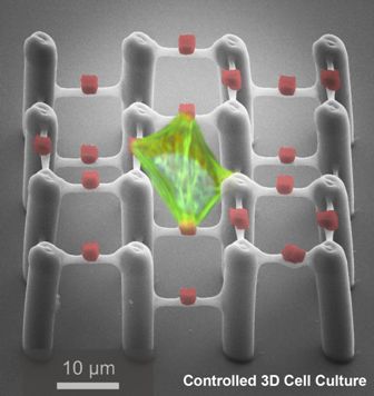
A cell in the two-component polymer scaffold.
The photo composition is based on a scanning electron microscopy and
laser scanning microscopy. (Image: CFN)
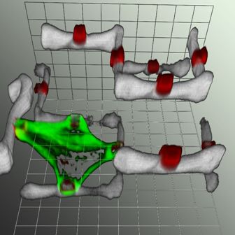
Laser-scanning microscopy (LSM) of the cell in
the two-component polymer scaffold. The cytoskeleton of the cell is
colored green, parts of the two-component polymer scaffold are
colored white, the “cell holds” are colored red. (Image: CFN)
The team used a special polymer scaffold consisting of a flexible, protein-repellent polymer with small box-shaped anchors made of a protein-binding material. For scaffold construction, the scientists used the Direct Laser Writing Method (DLS) developed by the physicists Professor Martin Wegener and Professor Georg von Freymann at CFN.
The scaffold consists of 25 µm high pillars that are connected by thin bars at various heights. In a second lithography step, the holds were placed exactly in the middle of the bars. With the help of a solution of adhesion proteins, the proteins only bind to these small holds. Within two hours, individual cells colonize the scaffolds and adhere to the given adhesion points only.
The research is an important step towards the general understanding of how the natural three-dimensional environment in the tissue influences the behaviour of cells.
Reference
Klein F, Richter B, Striebel T, Franz CM, Freymann Gv, Wegener M, and Bastmeyer M. Two-Component Polymer Scaffolds for Controlled Three-dimensional Cell Culture. Advanced Materials, Volume 23, Issue 11, pages 1341–5, March 18, 2011, DOI: 10.1002/adma.201004060