Microfluidic device separates non-spherical bioparticles for rapid disease diagnosis
23 April 2013
A team at the National University of Singapore (NUS) has developed a novel microfluidic device that separates and detects non-spherical bioparticles such as pathogenic bacteria and malaria-infected red blood cells. It can potentially be used for rapid medical diagnostics and treatment.
Though the team is focusing mainly on the rapid separation and detection of bacteria from pathological samples at the moment, their device has potential as a rapid diagnostic tool as well and could potentially replace an age-old method of detection based on bacterial culture.
Bioparticles such as bacteria and red blood cells (RBC) are non-spherical. Many are also deformable, for example, blood cells may change shape when affected by different pathogens in the body. Hence, the team’s shape-sensitive technique is a significant discovery. Currently, separation techniques are mostly designed for spherical particles.
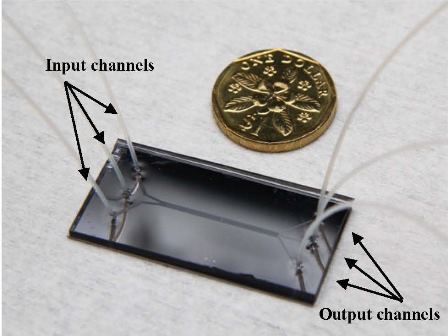
The microfluidic chip is only slightly
bigger than a Singapore $1 coin.
Source: National University of
Singapore
Project leader Assoc Prof Zhang Yong said, “The old method was developed about 100 years ago, but it is still being used today as the mainstream technique because no new technique is available for effective separation of bacteria from pathological samples like blood. Many of the pathogenic bacteria are non-spherical but most of microfluidic devices today are for separating spherical cells. Our method uses a special I-shape pillar array which is capable of separating non-spherical or irregularly-shaped bioparticles.”
The method developed by the NUS team can complete the diagnosis process in less than an hour compared to 24-48 hours required for bacterial detection by using conventional methods. Their device is also efficient in separating red blood cells (RBCs) from blood samples as RBCs are non-spherical. This enables rapid detection of diagnostic biomarkers which reside in blood sample.
One of the most challenging aspects for the team was designing and fabricating a device that is capable of detecting even the smallest dimension of bioparticles and still provide reasonably good throughput.
How it works and moving forward
Scientists have tried to address the problem of separating non-spherical bioparticles by using techniques such as restricting the flow of particles but these have not shown to be as effective. However, the NUS Bioengineering team’s I-shape pillar array device has proven to be successful.
The I-shape pillar array induces rotational movements of the non-spherical particles which in turn increases the effective hydrodynamic size of the bioparticles flowing in the device, allowing for efficient separation. Their design is able to provide 100 percent separation of RBCs from blood samples, outperforming conventional cylindrical pillar array designs.
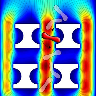
How the I-shape pillar array works.
Non-spherical cells such as rod-shaped ones are rotated by I-shape
pillar to increase their effective hydrodynamic size, isolating them
from samples. Source: National University of Singapore
The device can also potentially separate bioparticles with diverse shapes and sizes. The team has tested their device successfully on rod-shaped bacteria such as Escherichia coli (common bacteria which can cause food poisoning). So far, this has been difficult to achieve using conventional microfluidic chips.
Said Assoc Prof Zhang, “With our current findings, we hope to move on to separate other non-spherical bioparticles like fungi, with higher throughput and efficiency, circumventing the spherical size dependency of current techniques.”
The team’s findings were published in Nature Communications on 27 March 2013, titled: Rotational separation of non-spherical bioparticles using I-shaped pillar arrays in a microfluidic device.