IBM develops microfluidic probe to diagnose cancer in tissue samples
28 October 2013
IBM scientists are collaborating with pathologists at the University Hospital Zürich to test a prototype device called a microfluidic probe for accurately diagnosing different types of cancer.
The probe can detect molecular variations in cancerous tissue samples at the micrometer scale. The eight-millimeter-wide, diamond-shaped probe consists in its simplest form of a silicon microfluidic head ending with a small tip bearing two microchannels. These are used to finely control the quantity of reagents used to stain tissue.
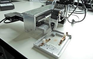
The prototype IBM microfluidic probe
A critical step in the diagnosis of cancer is the analysis of a patient's biopsy tissue sample, which sometimes can be as small as a pinhead. Even with such a small sample, pathologists can test for the absence or presence of tumour cells and provide important information pertaining to the course of treatment to doctors.
To analyze samples, pathologists typically stain the tissue sample with liquid reagents. The intensity and distribution of the colour stain classify and determine the extent of the disease. While this approach provides insights into the tumour, it is increasingly being realized that significant variations exist within the tumour itself; mapping these variations may help understand the drivers for each tumour, and consequently assist in personalizing treatment strategies.
“Pathologists are determined to obtain as much accurate information as possible from markedly small biopsy samples”, said Prof. Dr. Alex Soltermann, a pathologist specializing in lung cancer at the Institute for Surgical Pathology of the University Hospital Zürich.
“We hope to introduce new technologies, such as the microfluidic probe, into the clinical molecular pathology diagnostic framework to enable a range of investigations, which were previously thought to be infeasible. If we are successful, the tool will be a driver for personalized medicine, and translate into increased confidence in diagnosis and better detection of predictive cancer markers.”
Privatdozent Dr Peter Schraml, director of the tissue biobank at the Institute of Surgical Pathology, University Hospital Zürich, said, “In addition to assisting in diagnostics, this tool may provide insight into the biomarker distribution in tumour tissues, which can aid in understanding cancer progression.”
“For about a year we have been testing the probe in our lab, and initial results are very encouraging. We are now developing the technology in the context of important aspects in pathology” said Dr. Govind Kaigala, a scientist at IBM Research, Zurich. “Over the next several months, we will install a prototype device at the hospital and work alongside pathologists.”
The tool which houses the microfluidic probe was recently made significantly more compact and user-friendly and today is roughly the size of a tissue box. It is now at stage where it may assist in studying the distribution of low numbers of cancer cells in biopsied samples.
How does it work?
The probe injects very small volumes of reagents on the tissue surface and then continuously aspirates the reagents to prevent spreading and accumulation. This approach is used to deliver and retrieve reagents locally in selected areas of a tissue section with pinpoint accuracy. This local interaction with the tissue sample helps in mapping the heterogeneity in the tissue.
“We are very excited to partner with IBM on the microfluidic probe technology to develop techniques for its use in the clinical pathology framework – this is a fine example of a translational research that could also help answer some basic science questions” says Prof. Holger Moch, head of the Institute of Surgical Pathology at the University Hospital Zürich.
IBM scientists aspire to eventually partner with a medical equipment manufacturer to license the technology and bring it to market as a tool to assist pathologists in making challenging and critical decisions. The microfluidic probes are designed and manufactured at the Binnig and Rohrer Nanotechnology Center on the campus of IBM Research, Zurich.