Prostate cancer diagnosed from tissue sample in 90 seconds
14 November 2014
The Fraunhofer Institute for Ceramic Technologies and Systems (IKTS) in Dresden has developed a device that can identify cancerous prostate tissue from a biopsy within one and a half minutes by using laser stimulated fluorescence.
Since the sample does not require a long preparation time and can be pushed directly into the device and analyzed directly after it has been taken, the patient does not have to wait for days after the biopsy in order to know the outcome. The doctor receives the results immediately and can talk with the patient much sooner about the next steps to take.
Dr Jörg Opitz, scientist at IKTS, describes the process, "The physician places the removed tissue sample on a base plate, slides it into the machine, presses a button — and within one and a half minutes, receives a reliable indication of whether the tissue in the sample is benign or malignant."
In the current procedure to determine if the change in the prostate is benign or due to a carcinoma, a sample of prostate tissue is taken from the patient by inserting a small needle into the prostate, using ultrasound images to assist with navigation. From this sample laboratory staff fabricate wafer-thin tissue sections — a laborious job that takes at least a day. Then the tissue sections are forwarded to a pathologist, who examines them under the microscope. Even for experienced pathologists it is often difficult to distinguish between benign and malignant tissue.
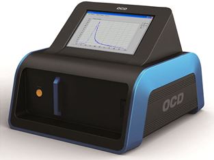
The prototype diagnostic device determines
whether the
prostate tissue sample is benign or malignant.
Source: Fraunhofer IKTS
Light stimulates the body’s own fluorescence
A further advantage of the new device is the reliability of the examinations. The analyses are based on autofluorescence of human tissue. There are 'fluorophore' molecules in every human body that re-emit light when a certain type of light falls on them.
In the device, a laser pulse is used to excite the fluorophore molecules in the tissue sample. The way in which this fluorescence radiation decreases differs between benign and malignant tissue and the scientists at IKTS have been able to determine a clear threshold for this different behaviour. Thus, the device can quickly detect if the collected sample contains cancerous tissue.
Each tissue type has a fixed but unique value, so prostate tissue has a different value from other tissue. Currently, the device can only be used for prostate cancer, since the unit is only calibrated for this tissue.
The researchers’ goal is to determine the threshold values for other tissue types and to integrate them into the analysis software of the device. Then, to identify cancer the operator would only need to enter the appropriate tissue type from a drop-down menu.
The diagnostic device has already completed its first two clinical studies, and the third study is currently underway.